Fig. 2.
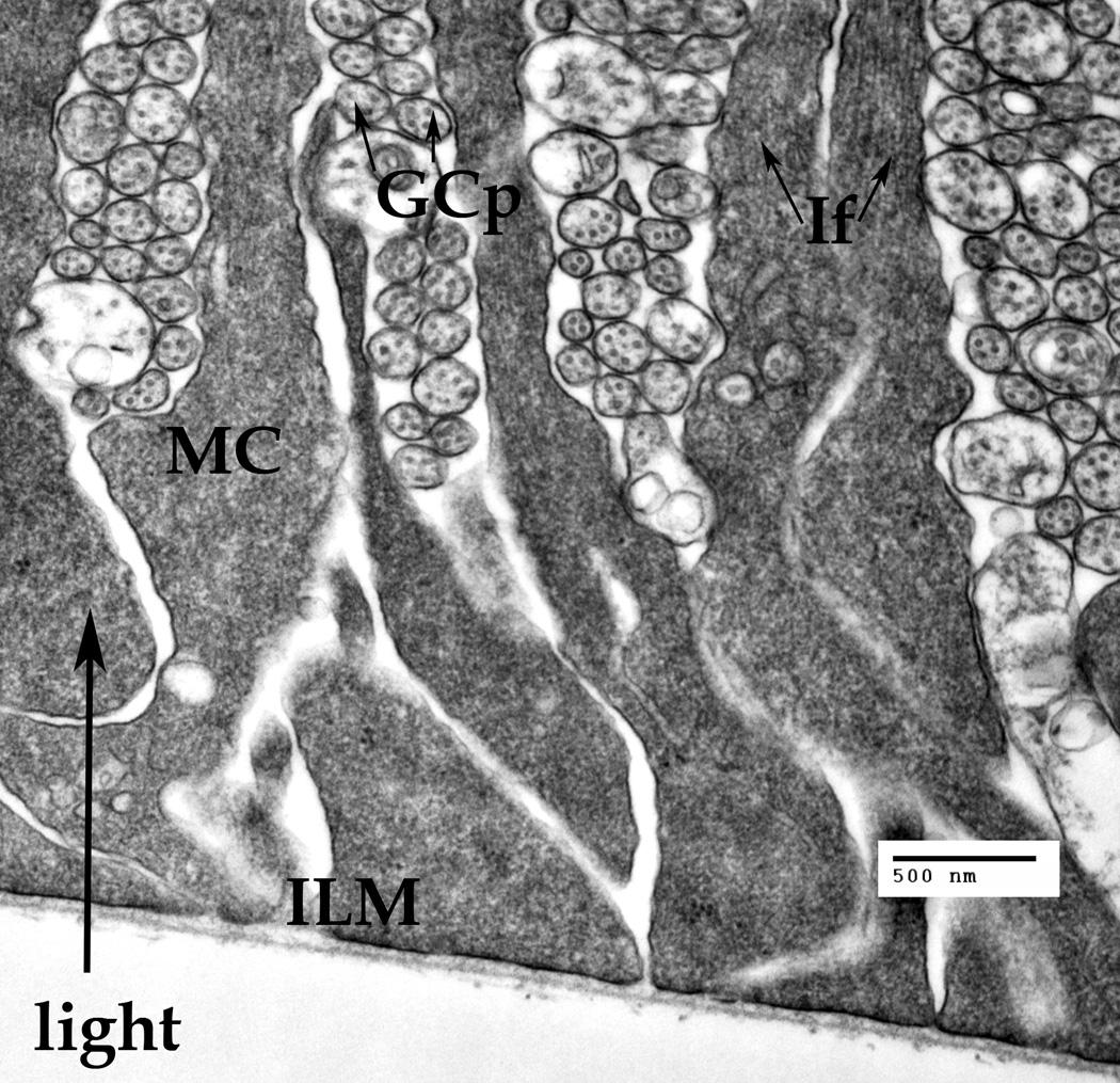
A cluster of the Müller cells' endfeet attached to the basal membrane thus forming the inner limiting membrane (ILM). The ganglion cell processes (GCp) are passing between the Müller cell endfeet. Cross-cut neuronal microtubules (arrows) are visible inside the GCps. The intermediate filaments (IF, arrows) are visible only in the narrow part of the Müller endfeet.
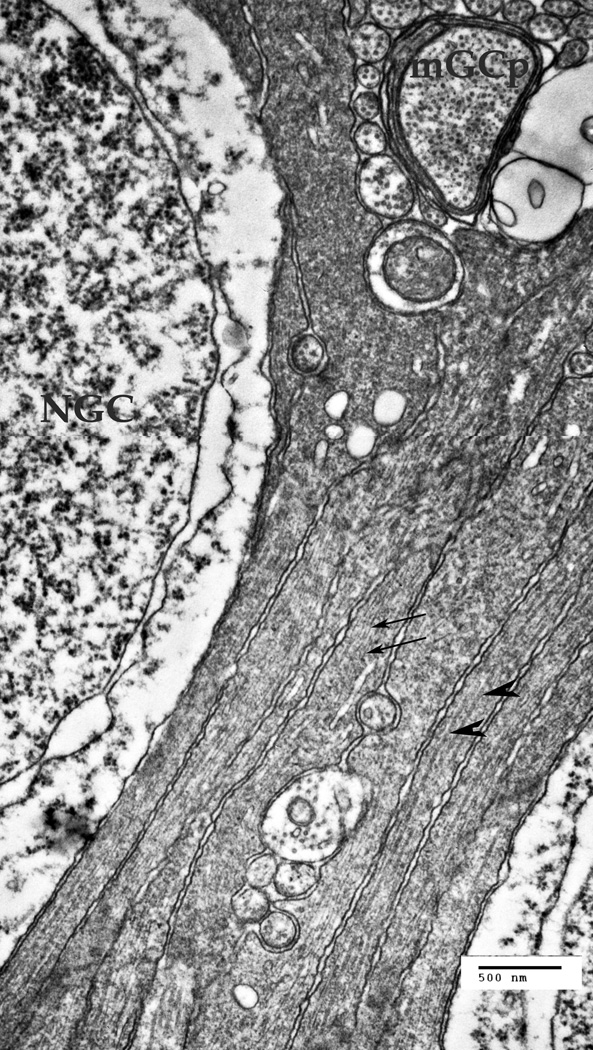
A. A cluster of the Müller cell processes at the ganglion cells' nuclear layer of the retina. NGC – the nucleus of a ganglion cell, mGCP – the myelinated process of a ganglion cell. The Müller cells processes are forced to go around other cells and their processes. Thin arrows point to the intermediate filaments in the MCs (10–18 nm), thick arrowheads point to microtubules (22–26 nm).
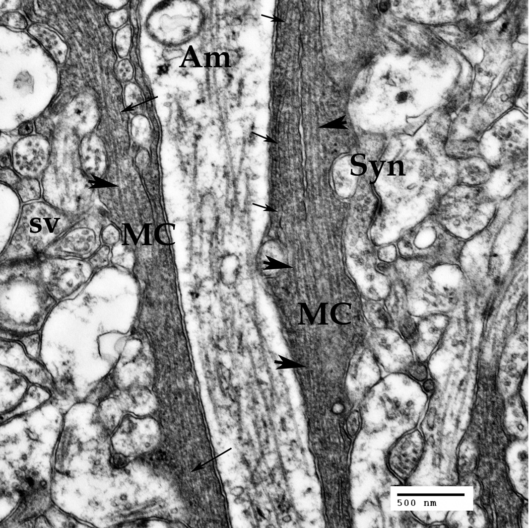
B. Müller cells' (MC) processes at the IPL level. A multitude of synapses (Syn) of ganglion, bipolar and amacrine (Am) neuronal cells are seen in this area. Synaptic vesicles (sv) are visible inside the synapses.
Thin arrows point to the intermediate filaments (10–18 nm), thick arrowheads point to microtubules (22–26 nm).
Bars: 500 nm


