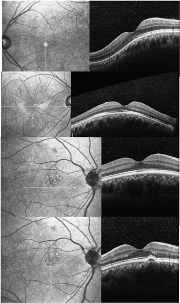
Figure 3.
Cross section enhanced depth imagining spectral domain OCT images with their orientation indicated by the thick straight lines lying on the left side fundi images. Top: vertically oriented dome-shaped maculopathy. Second from top: horizontally oriented dome-shaped maculopathy. Third from top and bottom: a bidirectional type dome-shaped maculopathy. A small juxtafoveal pigmented epithelial detachment is shown in the vertical orientation.

