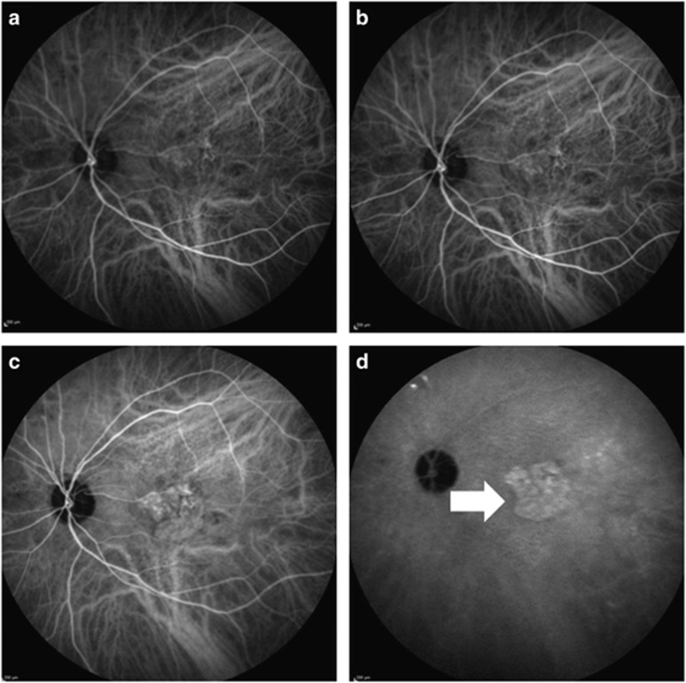
Figure 1.
The indocyanine green angiography images of a 78-year-old male neovascular age-related macular degeneration patient. (a and b). Early phases of angiography (c). The mid-phase of the angiography (d). The late phase of the angiography; white arrow shows the hot plaque formation secondary to occult choroidal neovascularization.

