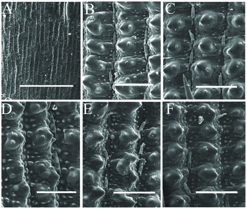
FIGURE 3.

Scanning electron microscopy observation of empty glumes, lemmas, and paleas. Epidermis of one empty glume (A), one lemma (B), and one palea (C) of wild- type plants. Epidermis of one empty glume (D), one lemma (E), and one palea (F) of g1–6 plants. Bars = 50 μm.
