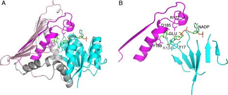
Figure 3.

The two‐domain structure of AAOR. (A) The N‐terminal NADP binding domain is in cyan, and the C‐terminal α/β‐domain in grey and pink (the α‐helical part in grey and the β‐sheet part in pink). The first β‐strand and following polypeptide chain containing the short and the long central α‐helices are highlighted in magenta. (B) The simplification of the major structural elements of the Gfo/Idh/MocA protein fold (as in AAOR). NADP is shown as a stick model. The side chains for the residues in the five key sites are shown as stick models. Also a glucose ligand (GLU, a substrate for AAOR) is shown.
