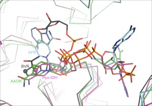
Figure 4.

The variation of the bound NAD(P) cofactor in the crystal structures of Gfo/Idh/MocA proteins. Different colors have been used for different cofactor conformations. NADP in AAOR is shown as green stick model, NADP in AFR in cyan, NAD in myo‐IDH, in purple, and NAD in RVB in grey. The C4 in the nicotinamide ring in every cofactor has been highlighted in spherical atom.
