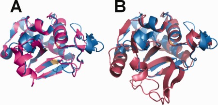
Figure 3.

Superposition of NlpC/P60 domains from MAP_1272c with non‐mycobacterial NlpC/P60 domain‐containing proteins. (A) Shown is superposition of MAP_1272c (blue) and B. cereus BCE_2878 (magenta) and (B) superposition of NlpC/P60 domains from MAP_1272c (blue) and D. vulgaris DVU_0896 (rust). Note that residues of the MAP_1272c catalytic triad are colored light orange for the purposes of orientation. The orientation of all structures is identical to that shown in Figure 2.
