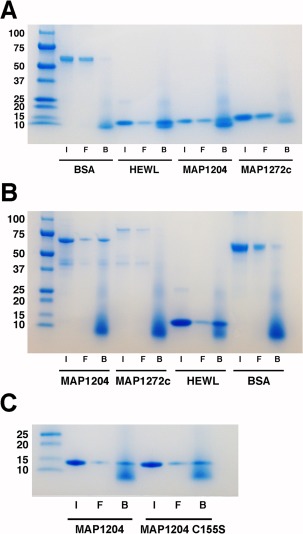
Figure 6.

SDS‐PAGE analysis of fractions from the peptidoglycan binding assay. MAP proteins without terminal tags are shown (A) along with positive (HEWL) and negative (BSA) controls. I = input protein, F = free/unbound protein from the supernatant, and B = bound protein from the pellet fraction. (B) MBP–MAP fusion proteins are shown along with positive (HEWL) and negative (BSA) controls. (C) The wild type and mutant (C155S) forms of MAP_1204 bound peptidoglycan in a similar manner. Protein size standards (in kDa) are at the left of each image.
