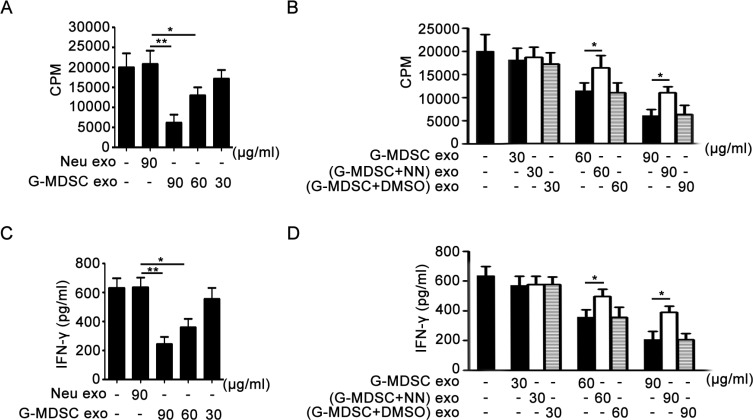
Figure 4. G-MDSC exo suppress CD4+ T cell proliferation and IFN-γ secretion correlating with Arg-1 activity in vitro.
(A and B). G-MDSC exo suppress CD4+ T cell proliferation through Arg-1 activity. CD4+ T cells were treated with G-MDSC exo (A) or/and (G-MDSC+NN) exo (B) at different concentrations for 72 h. The cultivation system was stimulated with anti-CD3 mAb (2 μg/ml) and anti-CD28 mAb (2 μg/ml). Cell proliferation was measured by [3H]-thymidine incorporation. (C and D). G-MDSC exo suppress IFN-γ secretion through Arg-1 activity. The levels of IFN-γ in culture supernatants from the CD4+ T cell proliferation system were detected using sandwich ELISAs. Data are shown as the mean ± SEM of each group (n = 6) pooled from three independent experiments. *p < 0.05, **p < 0.01, analyzed by ANOVA and Q test.

