
Figure 1. Representative images of different immune markers’ distribution in ESCC tissues detected with IHC staining.
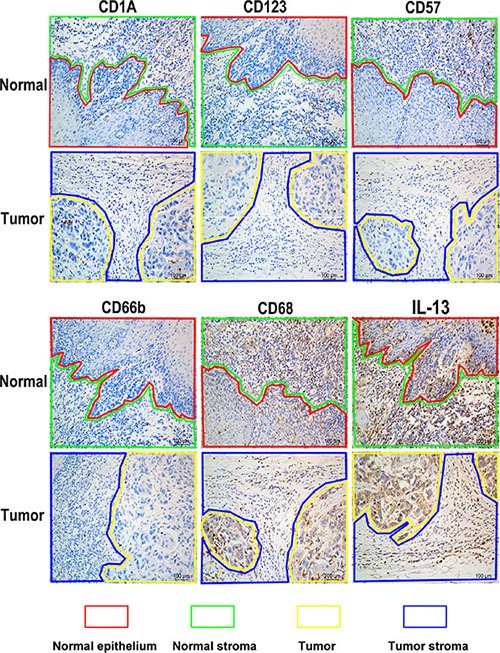
Lines of different color represented diverse areas of esophagus tissue. Positive stained cells were demonstrated as brown.
Official websites use .gov
A
.gov website belongs to an official
government organization in the United States.
Secure .gov websites use HTTPS
A lock (
) or https:// means you've safely
connected to the .gov website. Share sensitive
information only on official, secure websites.
Lines of different color represented diverse areas of esophagus tissue. Positive stained cells were demonstrated as brown.