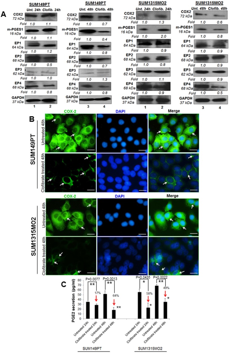
Figure 5. Effect of clofibrate treatment on the cyclooxygenase pathway.
Lysates prepared from A. SUM149PT and SUM1315MO2 cells untreated or treated with 20 μM clofibrate for 24 h and 48 h were Western blotted for COX-2, mPGES-1, EP1, EP2, EP3, EP4 and then stripped and re-probed with anti-GAPDH antibody to confirm equal loading. B. COX-2 immunostaining in SUM149PT and SUM1315MO2. SUM149PT and SUM1315MO2 cells were untreated or treated with 20 μM clofibrate for 48 h in eight-well chamber slides and then collected, permeabilized, and stained with an anti-COX-2 monoclonal antibody. Magnification, 40X. DAPI (Blue) was used as a nuclear stain and merged with COX-2 staining. Scale bar = 20 μm. C. Effect of clofibrate treatment on PGE2 secretion. Cell free culture supernatants of SUM149PT and SUM1315MO2 untreated or treated with 20 μM clofibrate for 24 h and 48 h were used to measure PGE2. Percent inhibition of PGE2 secretion was calculated by considering the secretion from untreated cells as 100%. * denotes statistically significant and ** represents statistically highly significant.

