
Figure 4. GBT promotes c-Myc degradation.
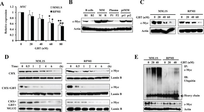
A. qPCR analysis for c-MYC expression in MM cell lines; B. Western blotting analysis showing the dose-dependent effect of GBT on c-Myc protein levels in MM cells; B cells, plasma, and MM cells from patients; C. Accelerated degradation of c-Myc protein after GBT treatment in MM.1S and RPMI8226 cells; D. MM.1S and RPMI8226 cells were treated with 50 nM of GBT, 20 μM of CHX, or the combination of GBT+CHX with 0.5 μM of MG132 for 6 h; E. Immunoprecipitation assay showing enhanced ubiquitination of c-Myc in MM.1S and RPMI8226 cells after GBT treatment. Aliquots from the above groups were directly analyzed by Western blot to test for c-Myc protein (bottom panel) in each group. Data represent mean ± SEM from three independent experiments. Statistical significances at **p<0.005 and *p<0.05 vs. respective vehicle/mock.