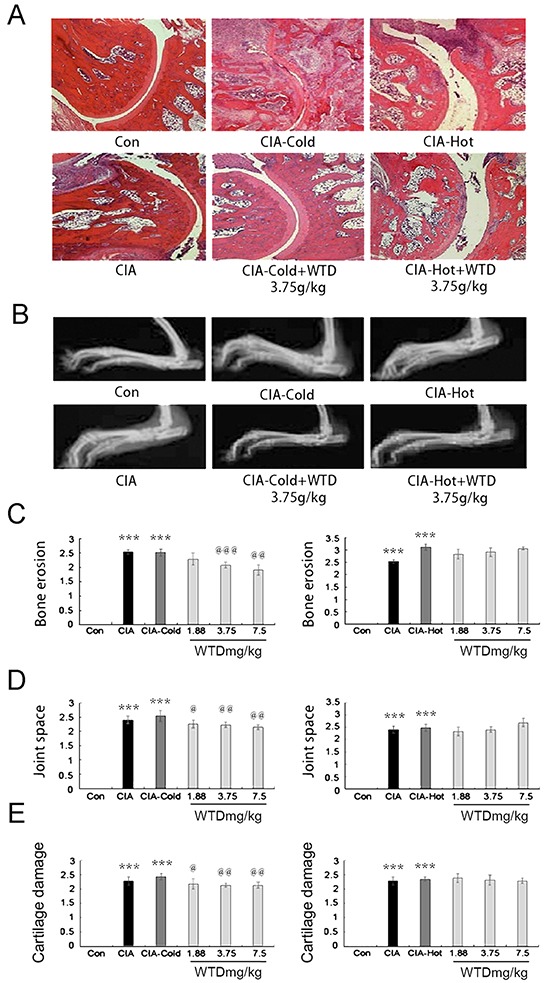
Figure 7. Effect of WTD on radiological changes and histologic lesions in CIA rats.

A. histologic observations of the joints in rats (HE staining); B. displays the clinical manifestation of CIA rats on day 21 after immunization, doses of 3.75 g/(kg•day) WTD improved paw swelling in the CIA-cold/hot model groups; C. D. and E. bone erosion, joint space and the degree of cartilage damage in joints, respectively, as described in the methods section. Data are represented as the mean±S.D (n=16). *, **, and ***, P<0.05, P<0.01, and P<0.001, comparison with the control group. #, ##, ###, P<0.05, P<0.01, and P<0.001, comparison with the CIA model group. @, @@, @@@, P<0.05, P<0.01, and P<0.001, comparison with the CIA-cold/hot model groups.
