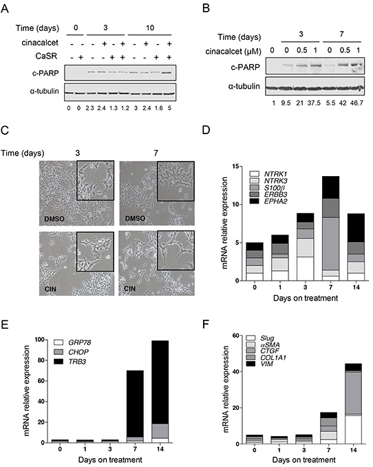
Figure 3. Prolonged in vitro exposure to cinacalcet induces apoptosis and cytodifferentiation in surviving neuroblastoma cells.

A. CaSR-positive and -negative SK-N-LP cells were grown in the presence of 1 μM cinacalcet or DMSO. Cells were lysed at indicated times to perform immunoblots. Bands intensity was quantified relative to that of α-tubulin. Blots shown are representative of three independent experiments. B. LA-N-1 cells were grown in the presence of indicated doses of cinacalcet or DMSO. Cells were lysed at indicated times to perform immunoblots. Data shown are representative of three independent experiments. C. Microphotography of LA-N-1 cells grown in the presence of 1 μM cinacalcet or DMSO for 3 and 7 days. D. Total RNA was isolated from LA-N-1 cells exposed to 1 μM cinacalcet or DMSO at indicated days. Gene expression analyses were carried out by RT-qPCR. Transcript levels were quantified relative to those detected in cells exposed to DMSO for the same number of days, and compared to time 0 (cells collected 16 hours after plating). Graph shows markers involved in neuroblastoma differentiation. E. Graph showing relative expression levels of transcripts involved in ER stress in cells processed as in panel D. F. Plot of transcripts involved in EMT analyzed as in panel D. See also Table 1 to compare gene expression patterns in three cell lines exposed to 1 μM cinacalcet or DMSO for 14 days. Data showed in panels C, D, E and F are representative of at least two independent experiments for each cell line.
