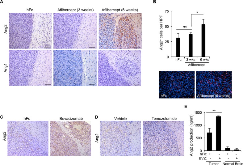
Figure 1. Anti-VEGF therapy-induced invasive tumor phenotype is associated with increased Ang2 expression.
(A) Sections of U87MG-derived tumors from mice treated with aflibercept for 3 weeks or 6 weeks or with control treatment (hFc) were stained for Ang2 and Ang1 expression. Invasive features and increased Ang2 were observed in animals treated with aflibercept for 6 weeks. Scale bars = 50 μm. (B) Quantification (top) of Ang2+ cells in tumors from animals treated with aflibercept (3 or 6 weeks) or control. Data are presented as mean ± SD. Representative images (bottom) show merged fluorescent Ang2 (red) and DAPI (blue). HPF, high-power field. ns, P > 0.05; *P < 0.05. (C, D) Tumor sections from mice treated with bevacizumab (C), temozolomide (D), or controls were stained for Ang2 expression. Scale bars = 50 μm. (E) Quantification by enzyme-linked immunosorbent assay of Ang2 production in tumor lysates from U87MG-derived intracranial xenografts after treatment with bevacizumab or control (hFc) compared with Ang2 present in normal brain tissue lysates. Data are presented as mean ± SD. BVZ, bevacizumab. **P < 0.01.

