
Figure 5. LATS1 suppressed the tumorigenicity and liver metastasis of SGC-7901 cells in vivo.
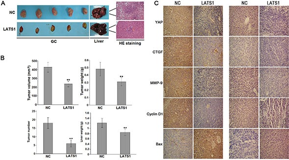
(A) Representative photographs of GC and metastatic liver tumors and HE staining of liver tissue derived from SGC-7901-LATS1 or SGC-7901-NC cells in SCID mice. (B) At the end of the experiment, the average volume and weight of gastric tumors in LATS1 group were significantly lower than those of the NC group. The number of metastatic liver tumor nodules and the average liver weight in LATS1 group were much lower than those of the NC group). (C) IHC analysis of the protein expression of YAP, CTGF, MMP-9, Bax and CyclinD1 in gastric tumors and metastatic liver tumors. The data were shown as the means ± SD of three independent experiments. **P < 0.01.