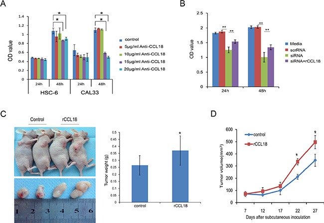
Figure 3. CCL18 promotes oral cancer cell growth in vitro and in vivo.

(A) OSCC cells (HSC-6 and CAL33) were treated with the indicated concentration of neutralizing CCL18 antibody (anti-CCL18). Cell viability was then measured using the CCK-8 assay at 24 h and 48 h. The data are presented as the mean ± SEM of triplicate experiments. (*P < 0.05) (B) HSC-6 cells were left untreated, transfected with 20 nM scrRNA, transfected with 20 nM siCCL18, or transfected with 20 nM siCCL18 and then treated with 20 ng/ml exogenous rCCL18 (siCCL18+rCCL18). Following treatment, cell viability was measured at 24 h and 48 h. The data are presented as the mean ± SEM of triplicate experiments (**P < 0.01). (C and D) Representative images of tumors obtained after subcutaneous injection of HSC6 cells into the flank region of nude mice, and treatment (3 times/week × 3 weeks) with vehicle control (n = 5) or exogenous rCCL18 (n = 6, 2 ng/g) starting on the 7th day. Tumor volumes were measured once every 5 days starting at the beginning of treatment. Four weeks after HSC-6 injection, the mice were sacrificed and the tumors were removed and weighed. Tumor weights (C) and volumes (D) are presented as the mean ± SEM. (*P < 0.05 vs. control).
