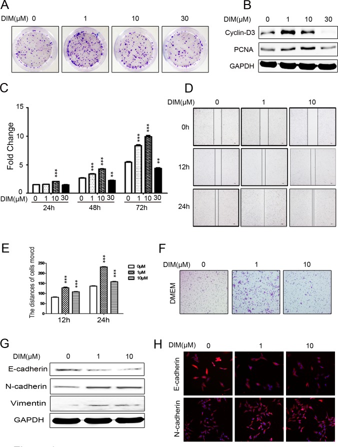
Figure 1. Low level of DIM promotes the proliferation and migration of gastric cancer cells.
A. Representative images of colony formation in HGC-27 cells treated with 0, 1, 10, and 30 μM DIM. Original magnification, 40 Χ. B. Western blotting assays for the expression of Cyclin-D3 and PCNA proteins in HGC-27 cells treated with 0, 1, 10, and 30 μM DIM for 48 h. C. Cell counting assay for HGC-27 cells treated with 0, 1, 10, and 30 μM DIM for 24, 48 and 72h. D. Wound healing assays for the migratory ability of HGC-27 cells at 12 and 24 h after the treatment with DIM (1 and 10 μM). Original magnification, 100Χ. E. The gap distance in A. was quantified. ***P < 0.001. F. The migratory ability of HGC-27 cells treated with 0, 1, and 10 μM DIM was evaluated by using transwell migration assay. Original magnification, 100 Χ. G. Western blotting assays for the expression of E-cadherin, N-cadherin, and Vimentin in HGC-27 cells treated with 0, 1, and 10 μM DIM for 48 h. H. Representative immunofluorescence images of E-cadherin and N-cadherin expression in HGC-27 cells treated with 0, 1, and 10 μM DIM for 48 h. Original magnification, 200 Χ.

