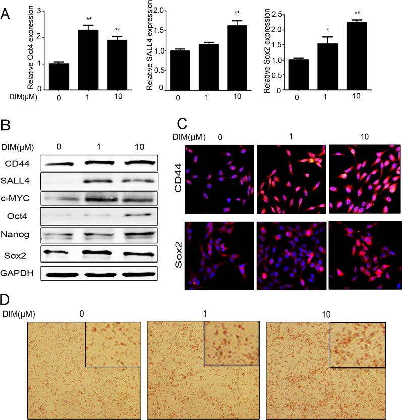
Figure 2. Low level of DIM enhances the stemness of gastric cancer cells.
A. Real-time RT-PCR for the expression of Oct4, SALL4, and Sox2 genes in HGC-27 cells treated with 0, 1, and 10 μM DIM for 48 h. (n = 3, P < 0.05). B. Western blotting assays for the expression of CD44, SALL4, c-MYC, Oct4, Nanog, and Sox2 proteins in HGC-27 cells treated with 0, 1, and 10 μM DIM for 48 h. C. Immunofluorescent staining of CD44 and Sox2 proteins in HGC-27 cells treated with 0, 1, and 10 μM DIM for 48 h. Original magnification, 200Χ. D. Adipogenic differentiation of HGC-27 cells treated with 0, 1, and 10 μM DIM for 48 h. Original magnification, 200 Χ.

