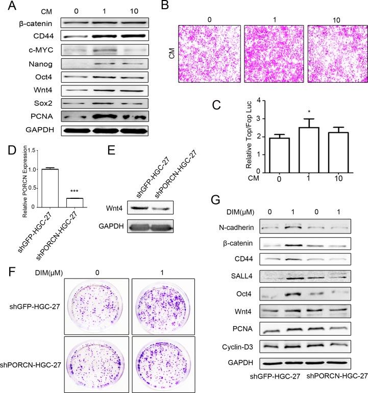
Figure 4. Low level of DIM activates β-catenin signaling by inducing Wnt autocrine.
A. Western blotting assays for the expression of β-catenin, CD44, c-MYC, Nanog, Oct4, Wnt4, Sox2, and PCNA proteins in HGC-27 cells incubated with the conditioned medium form DIM-treated HGC-27 cells for 48 h. B. The conditioned medium in the lower chamber of the transwell was collected from HGC-27 cells treated with 0, 1, and 10 μM DIM for 48 h. Original magnification, 100Χ. C. HEK293T cells transfected with the TOP-Flash or FOP-Flash luciferase reporter were incubated with equal amounts of conditioned medium collected form HGC-27 cells that were treated with indicated concentrations of DIM for 48 h. The ratio between TOP & FOP Flash luciferase activity was determined at 24h after treatment (n = 3, *P < 0.05). D. HGC-27 cells were transfected with PORCN-shRNA (shPORCN-HGC-27) or GFP-shRNA (shGFP-HGC-27) lentivirus. The expression of PORCN in HGC-27 cells was determined by real-time RT-PCR. (n = 3, ***P < 0.001). E. Western blotting assays for Wnt4 expression in shGFP-HGC-27 and shPORCN-HGC-27 cells. F. Representative images of colony formation in shGFP-HGC-27 and shPORCN-HGC-27 cells treated with 0 and 1μM DIM. Original magnification, 40Χ. G. Western blotting assays for the expression of N-cadherin, β-catenin, CD44, SALL4, Oct4, Wnt4, and PCNA proteins in shGFP-HGC-27 and shPORCN-HGC-27 cells treated with 0 or 1 μM DIM for 48 h.

