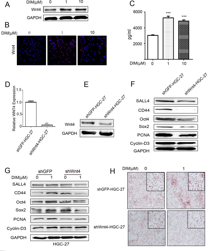
Figure 5. Low level of DIM enhances Wnt4 autocrine to activate β-catenin signaling.
A. Western blotting assays for Wnt4 expression in HGC-27 cells treated with 0, 1, and 10 μM DIM for 48 h. B. Immunofluorescence analyses of the expression of Wnt4 in HGC-27 cells treated with 0, 1, and 10 μM DIM for 48 h. Original magnification, 200Χ. C. ELISA assays for Wnt4 concentration in the conditioned medium from HGC-27 cells treated with low level of DIM for 48 h (n = 3, ***P < 0.001). D. HGC-27 were transfected with WNT4-shRNA (shWNT4-HGC-27) or GFP-shRNA(shGFP-HGC-27) lentivirus, respectively. The expression of WNT4 in HGC-27 cells was determined by using real-time RT-PCR. (n = 3, ***P < 0.001). E. Western blotting assays for Wnt4 expression in shGFP-HGC-27 and shWNT4-HGC-27 cells. F. Western blotting assays for the expression of SALL4, CD44, Oct4, Sox2, PCNA, and Cyclin-D3 proteins in shGFP-HGC-27 and shWNT4-HGC-27 cells. G. Western blotting assays for the expression of SALL4, CD44, Oct4, Sox2, PCNA, and Cyclin-D3 proteins in shGFP-HGC-27 and shWNT4-HGC-27 cells treated with or without 1μM DIM for 48 h. H. Adipogenic differentiation of shGFP-HGC-27 and shWNT4-HGC-27 cells treated with 0 and 1μM DIM for 48 h. Original magnification, 200Χ.

