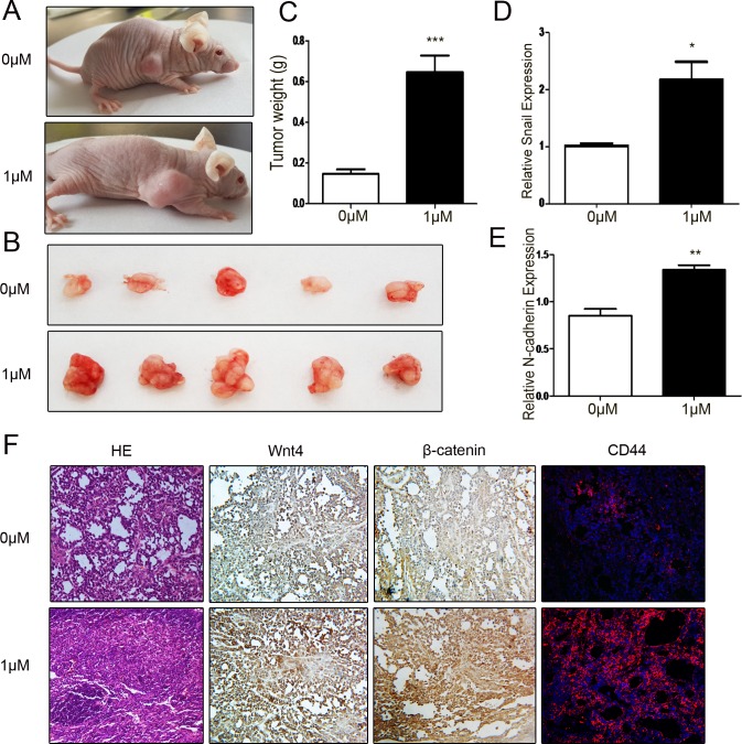
Figure 6. Low level of DIM promotes gastric cancer growth in vivo.
A. Representative images of tumor-bearing mice. B. The photographs of excised tumors at 28 days post-inoculation. C. Tumor weight was evaluated in mice transplanted with SGC-7901 cell that were treated with 0 or 1 μM DIM for 48 h (n = 5, ***P < 0.001). D. Real-time RT-PCR analyses of Snail and N-cadherin mRNA expression in the xenograft tumors. *P < 0.05, **P < 0.01. F. The subcutaneous tumors derived from SGC-7901 cells treated with 0 (upper) and 1 μM DIM (lower) was subjected to H&E staining, immunohistochemical staining of β-catenin and Wnt4, and immunofluorescent analyses of CD44 expression. Original magnification, 200×.

