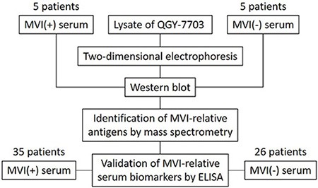
Figure 2. Schema of screening and validation of serum biomarkers for MVI.

The lysate of QGY-7703 is separated by two-dimensional electrophoresis followed by immunoblotting with the serum from 5 MVI (+) HCC patients or 5 MVI (−) HCC patients, respectively. The protein entities of spots that are either specifically associated with MVI (+) patients or with MVI (−) patients are identified by mass spectrometry. ELISA is performed to validate the presence of antibodies in the sera from 35 MVI (+) and 26 MVI (−) HCC patients that recognize MVI associated antigens.
