
Figure 3. Sero-proteomic identification of antigens that induce autoantibodies specifically in either MVI (+) or MVI (−) patients.
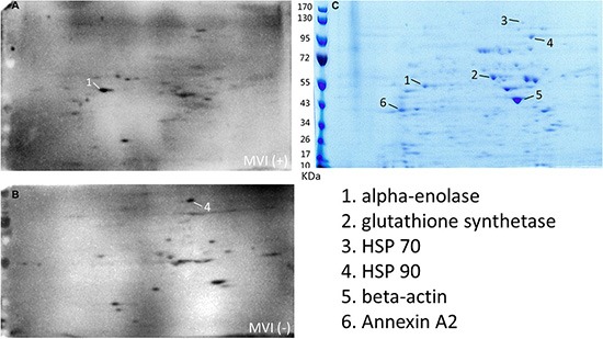
(A) Multiple identical two-dimensional electrophoresis gels were run to separate the HCC cell lysate and transferred to the membrane. Membranes were blotted with the sera from 5 MVI (+) HCC patients, respectively. One representative blot was shown. (B) Membranes were blotted with the sera from 5 MVI (−) HCC patients, respectively. One representative blot was shown. (C) One two-dimensional electrophoresis gel was stained with Coomassie brilliant blue. Six spots were repeatedly seen in either the blot with MVI (+) sera or the blot with MVI (−) sera, but not both.