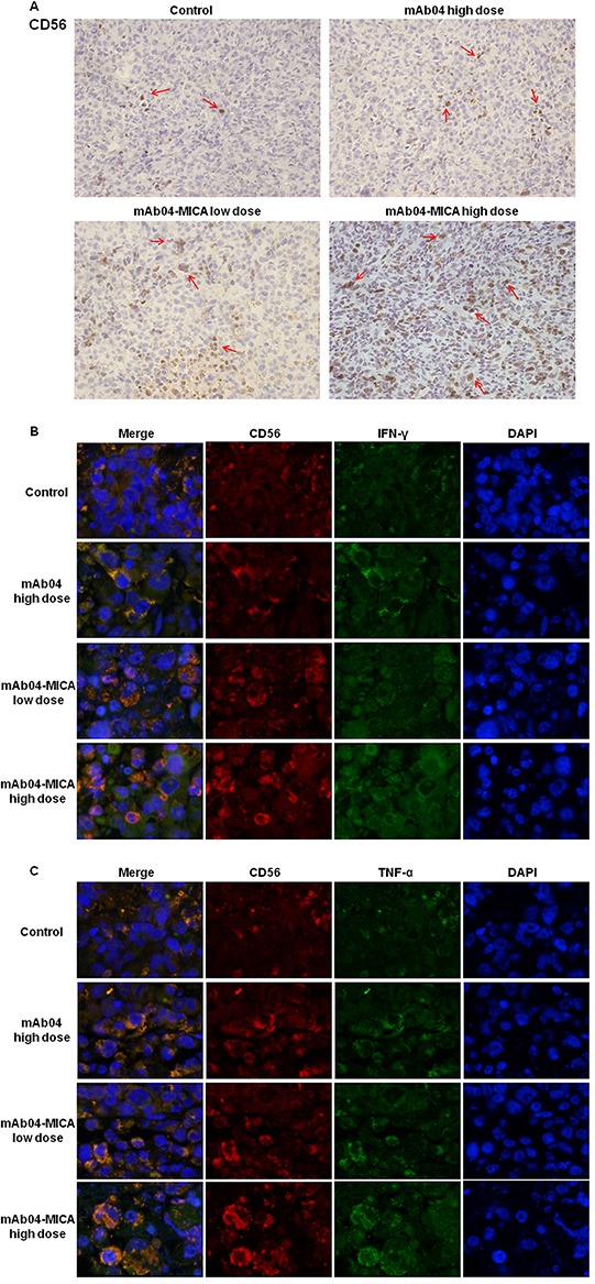
Figure 11. mAb04-MICA aggravated soakage of NK cells in MDA-MB-231 tumor tissue and increased the production of IFNγ and TNF-α by NK cells.

A. Infiltrated CD56+ cells were detected by IHC staining (brown staining, indicated by the red arrows) on serial sections, demonstrating more distribution of NK cells with mAb04-MICA treatment. B, C. IF double staining of CD56 (red fluorescence) and IFNγ/TNF-α (green fluorescence) to determine the expression level of IFNγ/TNF-α by NK cells. The orange staining cells after merged indicated the IFNγ/TNF-α expressing NK cells, which increased with the increase of dose.
