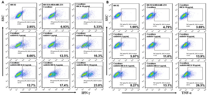
Figure 8. NK92 cells secreted more cytokines when treated with mAb04-MICA in the coculture with MDA-MB-231 cells.

A, B. Flow cytometry data represented the distribution of cytokine positive cells among NK92 cells, which indicated the proportion of NK92 cells expressing IFNγ/TNF-α along the x-axis increased as the treatments concentration increased. The percentage of IFNγ/TNF-α positive cells was calculated by FlowJo software.
