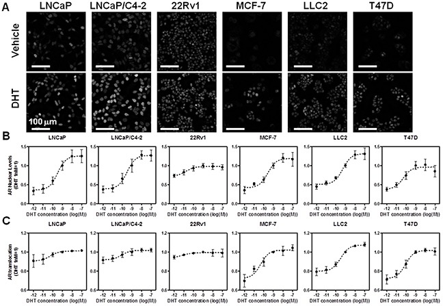
Figure 1. High throughput microscopy-based analysis of endogenous AR nuclear level and translocation across prostate and breast cancer cell lines.

A. Representative images of the cell lines used in HTM labeled with AR441 antibody after 24 hrs of treatment with either vehicle (top) or 1 nM DHT (bottom). B–C. DHT six point dose response and analysis of AR nuclear levels (B), or translocation (C), as determined by HTM and image analysis protocols
