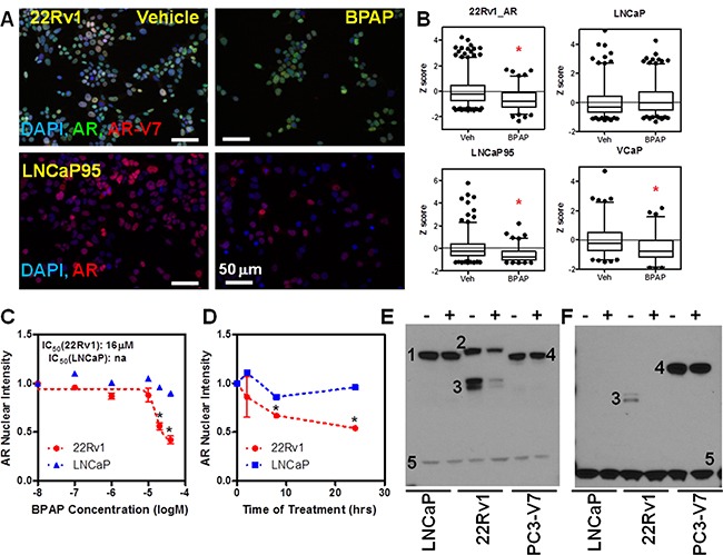
Figure 4. BPAP reduces AR full length and AR-V7 protein level in CRPC cell models.

A. Representative immunofluorescence images showing total AR and AR-V7 nuclear levels in 22Rv1 (A, top) and total AR in LNCaP95 (A, bottom) cells treated with vehicle or 20μM BPAP for 24 hrs. B. Single cell analysis of AR nuclear levels in 22Rv1, LNCaP95, VCaP and LNCaP (>1000 cells/condition) represented as box plot. *p<0.01 vs. vehicle. C–D. BPAP dose response and time course analysis in 22Rv1 cells by AR IF. E–F. Western blot in LNCaP, 22Rv1 and GFP-AR-V7:PC3 using AR (E) and AR-V-7 (F) antibodies showing reduction of AR (full length and variants) only in 22Rv1. Cells were treated with 20mM BPAP for 24 hrs. In the Western blots, 1 indicates full length AR, 2 full length AR with DBD duplication that is expressed in 22Rv1, 3 AR variants (AR-V7 in F), 4 GFP-AR-V7, 5 GAPDH.
