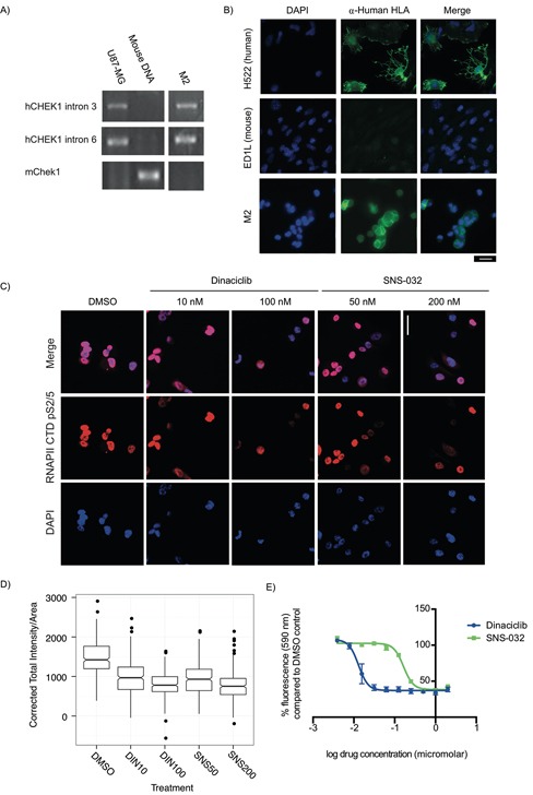
Figure 4. Explant cells generated from PDX models for rapid testing of targeted therapeutics are sensitive to CDK9 inhibitors.

A-B. PCR primers specific to human CHEK1 and mouse Chek1 were used to identify mouse and human content in PDX explant cells, using U87-MG cells as a human positive control and mouse genomic DNA as a mouse positive control. (A) Cells from a liver metastasis explant (M2) had human CHEK1 DNA but did not have mouse Chek1 DNA. (B) To validate this, α-Human HLA class 1 A, B, and C expression (a marker of cells of human and not mouse origin) were examined by immunofluorescence. All M2 explant cells evaluated were found to be α-Human HLA A, B and/or C positive. Human (H522) and mouse (ED1L) cell lines were used as controls. Scale bar: 20 μm. C. A KRAS mutant PDX explant treated for 24 h with the CDK9 inhibitors dinaciclib (10 nM and 200 nM) and SNS-032 (50 nM and 200 nM) has reduced Ser2/5 phosphorylation of the CTD of the large subunit of RNAPII. Scale bar: 20 μm. D. Box plots of the quantification of total fluorescent intensity per unit area of cells treated with dinaciclib and SNS-032 and stained for p-Ser2/5 RNAPII CTD. Data presented was collected in three independent experiments. E. Dinaciclib (IC50: 13 nM) and SNS-032 (IC50: 165 nM) inhibit the growth of explant cells during a 3 day treatment. Data presented is the average fluorescence and corresponding standard error of the mean for three independent experiments.
