Fig. S1.
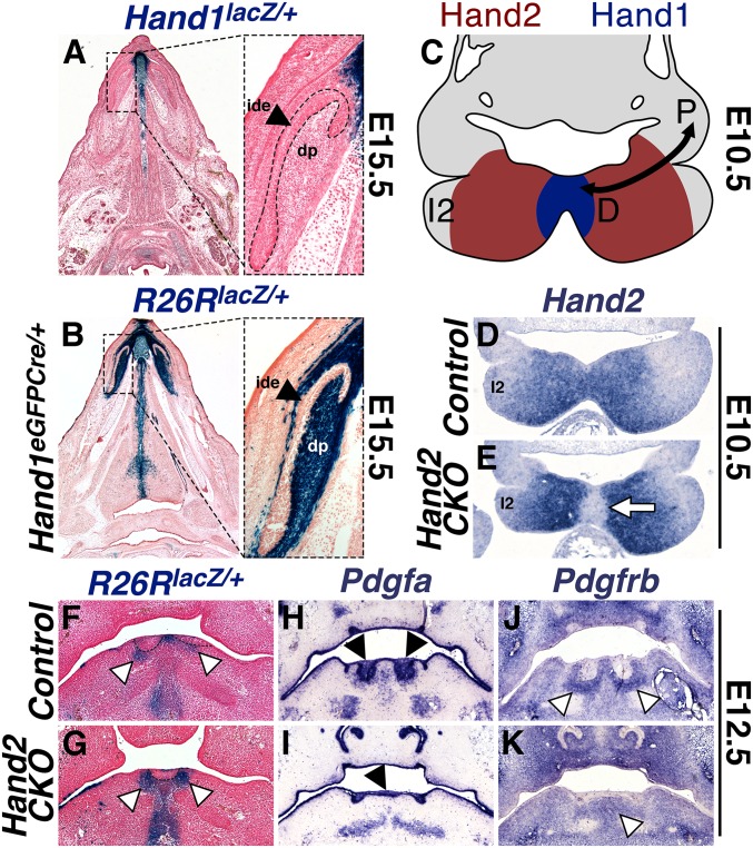
Hand1 (H1)-lineage cells form the ectomesenchymal component of the mandibular incisors in a Hand factor-dependent manner. (A) X-Gal staining of the H1lacZ knock-in allele reveals that, at E14.5, neither the dental epithelium (de) nor the dental mesenchyme (dm) of the mandibular incisor is positive for Hand1 expression. (B) X-Gal staining of the R26RlacZ reporter reveals that, at E15.5, the mandibular incisor dental papilla (dp) is composed of H1Cre knock-in allele lineage cells. Inner dental epithelium (ide) is marked with a black arrowhead. (C) H1 is expressed in the distal (D) cells of the mandibular arch (MD1; denoted by I2 in all figures), wherein it overlaps with Hand2 (H2) expression, although H2 expression extends more proximally (P). (D and E) In situ hybridization of E10.5 sections shows that the H1Cre conditionally ablates H2 expression within the crNCCs of the distal cap (white arrow), but not from the remainder of MD1. (F and G) Frontal sections of E12.5 embryos stained with X-Gal to visualize the R26RlacZ reporter reveals that two presumptive mandibular incisors are identifiable in Control embryos (outlined), whereas incisor development is arrested in Hand2 CKOs (H2fx/-;H1Cre/+). White arrowheads denote presumptive dp cells of the H1Cre-lineage. (H–K) In situ hybridization for the dental epithelial marker Pdgfα (black arrowheads) and the dental mesenchyme marker Pdgfrß (white arrowheads) (1, 2) in E12.5 embryos confirms the arrest of incisor development.

