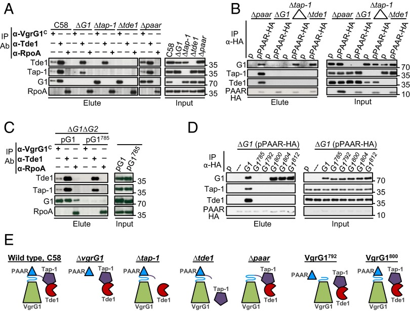
Fig. 7.
Interactions of VgrG1, Tap-1, Tde1, and PAAR proteins. Co-IP of WT A. tumefaciens C58 and indicated mutant strains (A) or ∆vgrG1∆vgrG2 (∆G1∆G2) expressing full-length vgrG1 (G1) and with the vgrG1 C31 deletion variant (G1785) (C) by using α-VgrG1 C-terminal epitope and α-Tde1 antibodies. α-RpoA antibody was used as a negative control. Co-IP of various mutants by using strains expressing pRL662 plasmid (p) only or PAAR-HA (B) or ∆G1(pPAAR-HA) expressing full-length VgrG1 (G1) or each of truncated VgrG1 variants (D) by anti-HA antibody. The resulting total protein extract was used as input for Co-IP. Coprecipitated proteins were detected by Western blot analysis with antiserum specific to indicated proteins. Proteins in input and elute fractions were detected by Western blot analysis. Protein names and molecular weight markers are indicated at the left and right, respectively. (E) A summary model of protein–protein interactions. Each protein is color-coded and C31 of VgrG1 is presented as β-helix consisting L5-β7 (L5-β5-L6 in blue and β6-L7-β7 in purple) at tip of VgrG spike.

