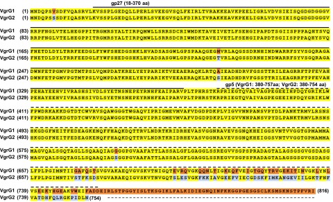
Fig. S1.
Amino acid sequence alignment of A. tumefaciens C58 VgrG1 and VgrG2. Identical residues are in yellow and variable residues in VgrG1 and VgrG2 are in orange and light blue, respectively. The solid and dashed lines represent regions with predicted gp27 and gp5 domains, respectively. Number in brackets at both sides of sequences is the residue number.

