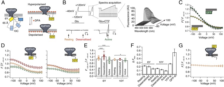
Fig. 5.
Voltage-dependent quenching of intracellular fluorophores by DPA. (A) Cartoon showing 90° arc of possible positions of the 10C site relative to 6Y, given a separation of 65 Å (from FLIM measurements). The scheme shows DPA translocation through the plasma membrane with hyper- and depolarizing voltage, and the change in distance to the 10Y insertion. (B) Schematic representation of the voltage ramp protocol used to drive DPA translocation across the membrane and the corresponding spectra acquisition. This protocol was used to record voltage-dependent fluorescence quenching by DPA at distinct time points during long glutamate stimulation (5 s) of cells expressing YFP-tagged GluA2. For assessment of quenching in different receptor states, the ramps were timed to translate DPA across the membrane in resting (yellow), desensitized (red), and active states. Fifty-millisecond frames were collected during each ramp in the absence and presence of CTZ. The corresponding fluorescence emission spectra were normalized to fluorescence intensity at −40 mV. AU, arbitrary units. (C) Voltage-dependent quenching of membrane-bound GFP (green circles) and YFP (yellow triangles). To eliminate hysteresis effects, data points are the average of responses to negative- and positive-going ramps. Lines represent weighted sigmoid fits. (D) Quenching of fluorescence of GluA2-6Y (Left) and GluA2-10Y (Right) in resting (orange circles), desensitized (red circles), and active (green circles) states. (E) Summary of quenching at the hyperpolarizing limit, normalized to quenching at −40 mV for GluA2-6Y and GluA2-10Y. An unpaired two-tailed Student’s t test gave P = 9.6E-9 for 6Y, desensitized vs. resting; P = 0.001 for 6Y, active vs. resting; and P = 0.008 for 10Y, active vs. resting. (F) Summary of extent of quenching for the I6 and I10 positions in the different functional states compared with membrane-bound YFP-tk and GFP-tk. The curve for the control position I0 was subtracted from all of the quenching curves before fitting to a sigmoid function. tk, truncated k-ras sequence (see Materials and Methods). (G) Lack of voltage dependence of GluA2-0Y.

