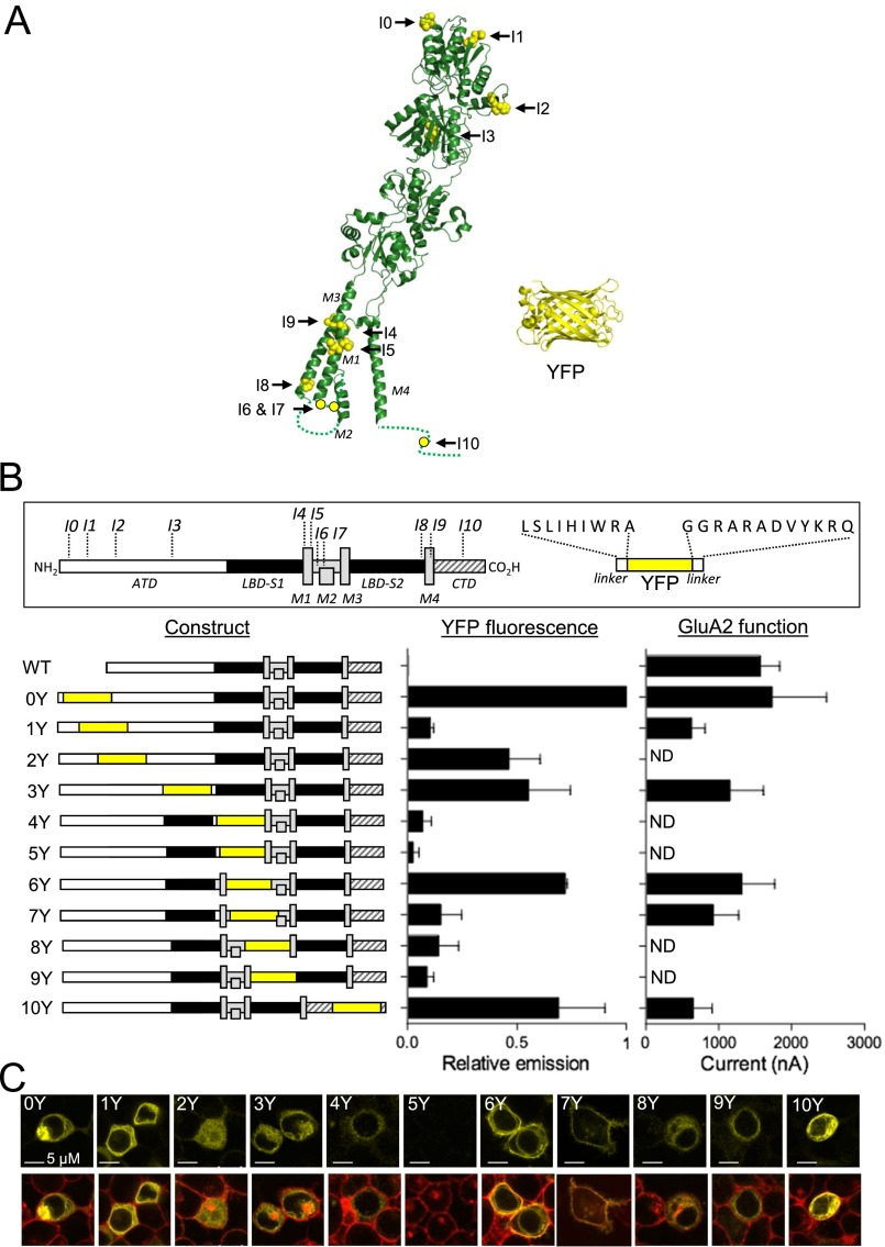
Fig. S1.
Insertion of YFP in the GluA2 subunit. (A) Structure of the GluA2 subunit [Protein Data Bank (PDB) ID code 3KG2] with positions of insertion points (I0 to I10) and the structure of YFP (PDB ID code 1F0B). (B) Schematic overview of GluA2 subunit topology and YFP insert (box) and summary of the 11 GluA2-YFP constructs (Left) and their relative fluorescent (Middle) and functional (Right) properties. YFP fluorescence intensities were measured in suspensions of HEK293T cells transfected with GluA2-YFP constructs at the emission maximum for YFP (530 nm) and are shown normalized to the intensity measured for GluA2-0Y. Data represent the mean ± SEM of three independent transfection experiments. Amplitudes of membrane currents evoked by glutamate (1 mM) in Xenopus oocytes expressing GluA2-YFP constructs at a membrane potential of −60 mV (Materials and Methods) are shown. Data represent the mean ± SEM for five to 12 oocytes. ND, not detected. (C) Confocal photomicrographs of HEK293T cells expressing GluA2-YFP constructs. YFP fluorescence (Upper, yellow) overlaid with images of DeepRed-stained cell surface membrane fluorescence (Lower, red). Colocalization of cell surface membrane stain and YFP fluorescence appears as orange.

