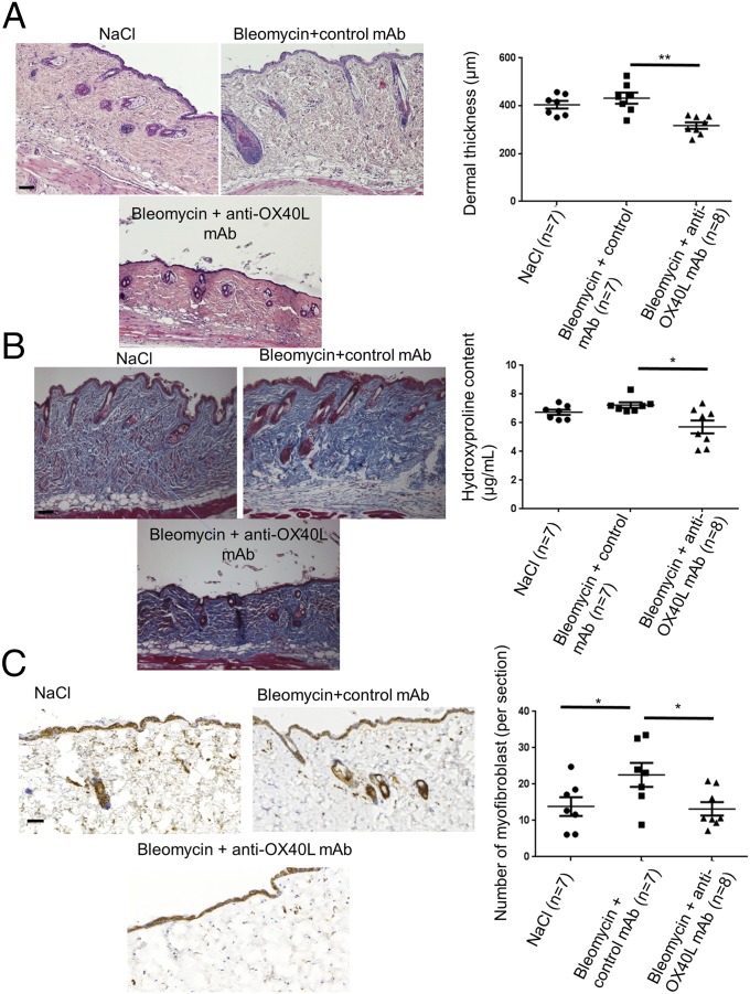
Fig. 6.
Inhibition of OX40L with a neutralizing mAb induces regression of established fibrosis in the bleomycin mouse model. (A) Representative H&E-stained sections (magnification: 100×) from C57/Bl6 mice injected s.c. with bleomycin for 6 wk and injected i.p. with IgG (control) for the last 3 wk (n = 7); C57/Bl6 mice injected s.c. with bleomycin for 6 wk and injected i.p. in parallel with anti-OX40L neutralizing mAb for the last 3 wk (n = 7); and C57/Bl6 mice injected s.c. with bleomycin for 3 wk followed by s.c. injections of NaCl for the last 3 wk (n = 8). (Scale bar: 100 µm.) (B) Representative sections stained by trichrome. (Magnification: 100×.) (Scale bar: 100 µm.) The hydroxyproline assay evaluates collagen content. (C) Myofibroblasts were identified by positive staining for α-SMA in slides after counterstaining with hemalun. (Scale bar: 100 µm.) Twenty-two mice were used for these experiments. Results are represented by dot blots with mean ± SEM. The experiment was performed in two independent series. *P < 0.05; **P < 0.01; two-sided Mann–Whitney test.

