Fig. 7.
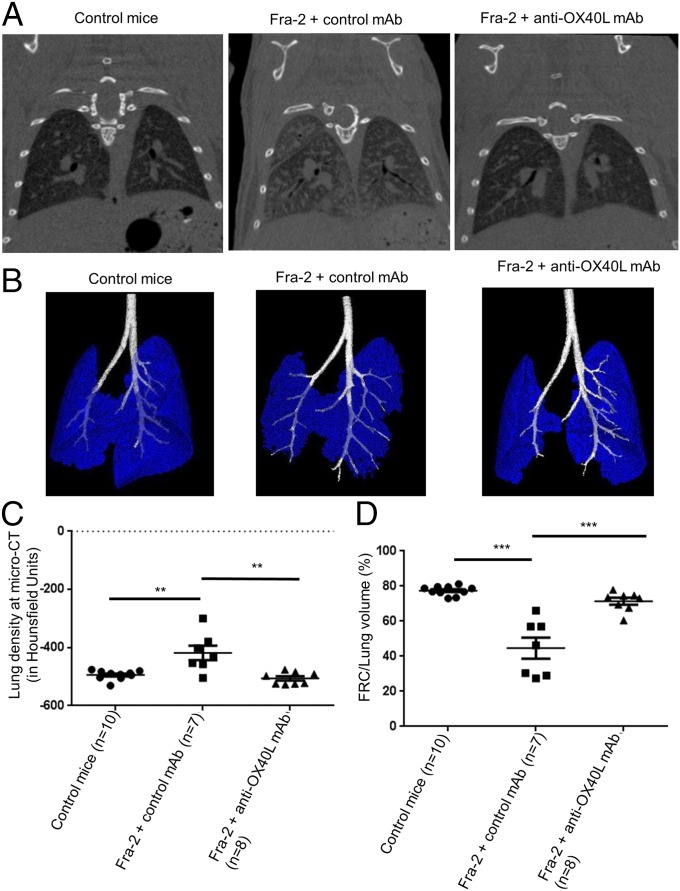
Inhibition of OX40L prevents the development of fibrosing alveolitis: CT-scan data. (A) Fibrosing alveolitis was observed in Fra-2 mice receiving control IgG. Representative micro-CT images are shown. (B) Representative images of functional residual capacity (in blue) in different mice; bronchi are in white. (C) Increased lung density at micro-CT in Fra-2 transgenic mice treated with control IgG (n = 7) compared with Fra-2 mice treated with anti-OX40L mAb (n = 8) or C57/BL6 wild-type (control) mice (n = 10). (D) Residual lung volume, expressed as the percentage of functional residual capacity on total lung volume. Values are dot blots with the mean ± SEM; **P < 0.01; ***P < 0.001; two-sided Mann–Whitney test.

