Fig. 9.
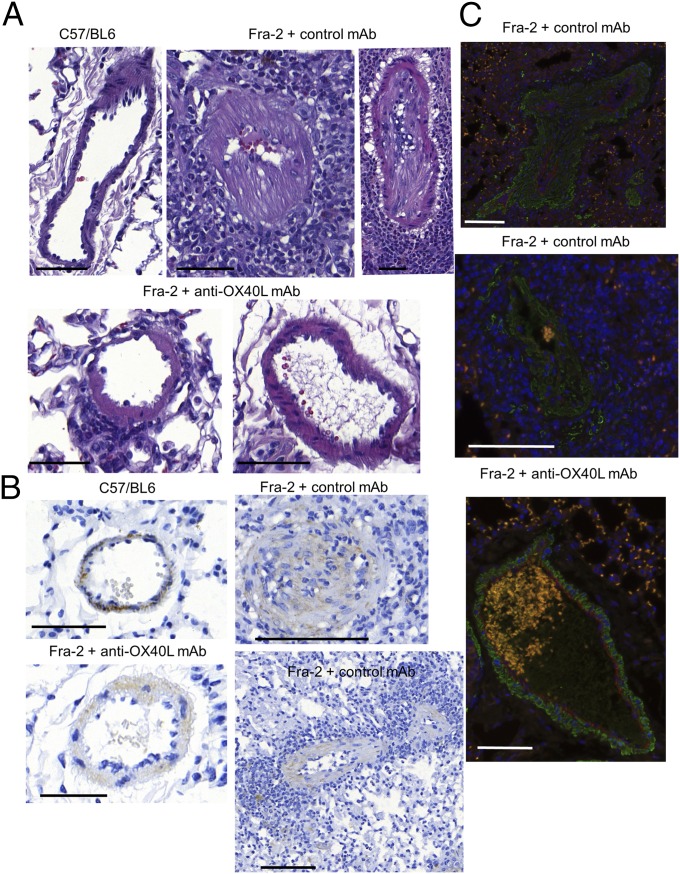
Inhibition of OX40L prevents PAH in Fra-2 mice. (A) Representative H&E-stained sections from Fra-2 mice treated with control antibody (two representative pictures are shown: one vessel is almost occluded, and one is completely occluded), from C57/BL6 (control) mice, and from Fra-2 mice receiving anti-OX40L antibody (two representative pictures are shown: one vessel with a mild infiltrate, and one vessel without vasculopathy or inflammatory infiltrate). (Scale bars: 100 µm.) (B) Remodeling of the vessel was assessed by staining for α-SMA (immunohistochemistry) and was more pronounced in Fra-2 mice receiving control IgG (vessel occluded or almost completely occluded with a massive inflammatory infiltrate on two representative pictures) than in Fra-2 mice treated with anti-OX40L mAb or C57/BL6 (control) mice. Twenty-five mice were used for this experiment. (Scale bars: 100 µm.) (C) There was no costaining for CD31 (red) or α-SMA (green) in vessels from Fra-2 mice treated with control antibody or anti-OX40L antibody. Vasculopathy in Fra-2 mice receiving control antibody was related to the prominent proliferation of fibroblasts (marked by α-SMA) rather than endothelial-to-mesenchymal transition (no coexpression of CD31 and α-SMA). A massive perivascular infiltrate was noticed (DAPI). (Scale bars: 100 µm.)

