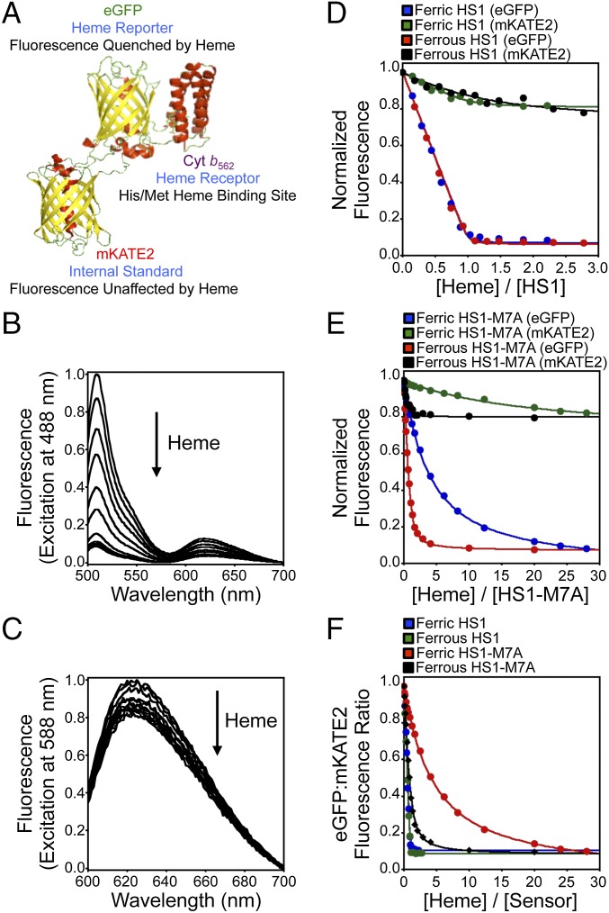
Fig. 1.
Design and heme-dependent fluorescence properties of the heme sensors. (A) Molecular model and design principles of the heme sensor, HS1. The model is derived from the X-ray structures of mKATE [Protein Data Bank (PDB) ID code 3BXB] and CG6 (PDB ID code 3U8P). Ferric heme-dependent changes in the normalized fluorescence emission spectra of HS1 at pH 8.0 upon excitation of EGFP (B, ex. = 488 nm) and mKATE2 (C, ex. = 588 nm) are illustrated. Normalized changes in EGFP (ex. = 488 nm, em. = 510 nm) and mKATE2 (ex. = 588 nm, em. = 620 nm) fluorescence upon titration of heme into 0.5 μM HS1 (D) and HS1-M7A (E) at pH 8.0 are illustrated. (F) Change in EGFP/mKATE2 fluorescence ratios for HS1 and HS1-M7A upon titration of heme. All titration data are fit to 1:1 heme/protein binding models.

