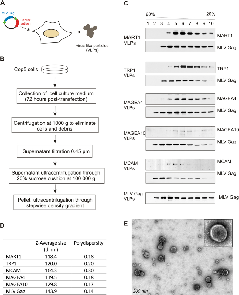
Figure 1. Generation and purification of VLPs.
(A) General workflow of generation of MLV Gag based VLPs. (B) Schematic representation of the purification protocol. (C) Western blot analysis of VLPs. Particles obtained with ultracentrifugation through 20% sucrose cushion were further centrifuged through stepwise sucrose density gradient (20%, 35%, 45%, 60%) at 120 000 g and 4 °C for 1.5 h in a Beckman SW55 rotor and divided into 10 fractions. The presence of VLPs in each fraction was analyzed by specific antibodies against melanoma antigens and the MLV Gag protein. (D) Physical characterization of VLPs ultracentrifuged through 20% sucrose cushion as assessed by DLS. The mean value of 4 × 10 measurements performed at 22 °C is shown. (E) Transmission electron micrograph of negatively stained MAGEA4 VLPs purified through density gradient centrifugation. Enlarged image of particle is shown on the upper-right corner of the picture.

