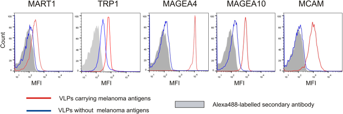
Figure 2. Surface analysis of melanoma antigens incorporated into VLPs.
FACS analysis was performed with VLPs bound to aldehyde/sulfate latex beads as described in Materials and Methods section. The red line corresponds to VLPs carrying recombinant melanoma antigens, the blue line to VLPs without cancer antigen, and grey area shows the signal obtained with secondary Alexa488-labelled antibody. One representative experiment out of the three performed is shown. MFI = Mean fluorescence intensity.

