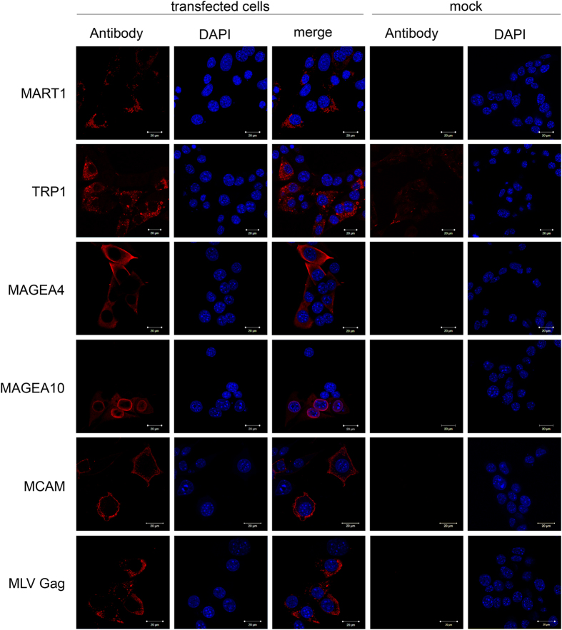
Figure 3. Subcellular localization of melanoma antigens used in this study.
The COP5 cells transfected with plasmids encoding for the MLV Gag protein and respective cancer antigen were grown on cover slips, fixed and incubated with specific antibodies and Alexa568-conjugated secondary antibody as described in Methods section. Untransfected cells (mock control) incubated with specific antibodies are shown. DAPI was used to stain nuclei of the cells.

