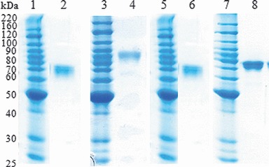
Figure 3.

Purification of hemagglutinin (HA) antigens from plant tissue. Purified HA antigens were analyzed by SDS–PAGE followed by Coomassie staining. Lane 1, 3, 5, and 7: Molecular marker, lane 2: HAB1(H1), lane 4 HAB1(H3), lane 6: HAF1(B), and lane 8: HAC1.
