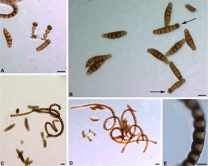
Fig. 11.

Pseudotorula helica. A. Conidiophores developed laterally on a hypha and each terminated by a corona cell (holotype). B. Mature phragmoconidia, arrows show corona cells at apex of two conidia (DAOM 96014e). C–D. Scolecoconidia illustrating the helical arrangement and the extensive melanization and periclinal thickening of the wall (holotype & DAOM 96014e). E. Enlargement of cells of a scolecoconidium illustrating periclinal thickening of the wall and melanization of the wall and septa (DAOM 96014e). Bars: A–D = 10 μm, E = 5 μm.
