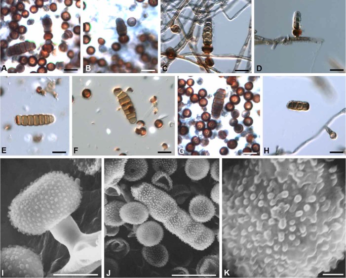
Fig. 2.

Bahusaganda elaeodes. A–D. Conidiogenesis illustrating corona cells and developing conidia (ILLS 36106). C–D. Corona cells laterally attached to hyphae (ILLS 36106). E–H. Mature conidia and corona cells (holotype). I. Corona cell and subtending stalk cell laterally attached to hypha (holotype). J. Mature conidium and corona cells (holotype). K. Conidium cell illustrating verrucae (holotype). Bars: A–H & J = 10 μm, I = 5 μm, K = 1 μm.
