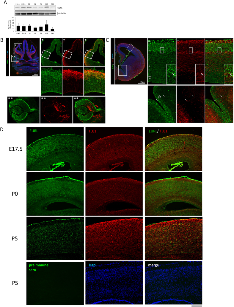
Figure 1. Expression of EURL during mouse brain development.
(A) Western blotting analysis of EURL protein levels (represented as an immunoreactive band of approximately 34kDa) in brain lysates over the course of mouse brain development, from embryonic (E14.5, E17.5) to postnatal (P0, P2, P5, P17, P30). Signals for β-tubulin (approximately 50kDa in size) serve as loading control. (B) Immunostaining reveals prominent EURL reactivity (green signal) within the dorsal telencephalon (region magnified in “*”) as well as the developing retina and lens (region magnified in “**”). Double immunostaining with the early neuronal marker TUJ1 (red) reveals EURL immunoreactivity in neural and non-neural cells. (C) Within the E14.5 dorsal telencephalon, EURL immunoreactivity is strong in posmitotic cells of the cortical plate (CP) and marginal zone (MZ), with a weaker signal detected in cells of the intermediate zone (IZ) and germinal subventricular/ventricular zone (SVZ/VZ). A CP neuron is represented with an arrow in the boxed insert. (D) At late embryonic (E17.5) stages, EURL immunoreactivity persists in CP neurons, as well as in postnatal (P0, P5) brains. The immunostaining signals detected with the EURL antibody are not evident when a parallel experiment is conducted with preimmune serum. Scale bar, 200 μm.

