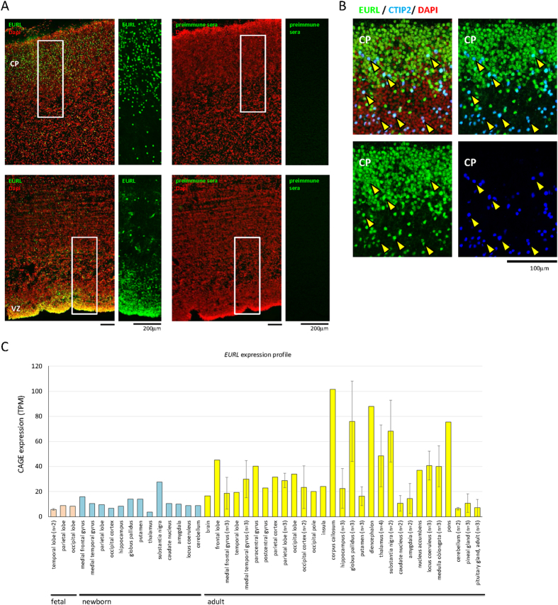
Figure 2. Immunostaining for EURL expression within the human fetal brain.
(A) Immunostaining of human fetal cerebral cortex at 16 weeks post conception (GW16) revealed prominent EURL immunoreactivity in cells of the germinal ventricular zone (VZ) and the developing cortical plate (CP). Parallel experiments with preimmune serum did not elicit a signal. (B) Co-labelling studies demonstrate that EURL is expressed in projection neurons marked with CTIP2 (arrowheads) Nuclei are stained with DAPI. (C) A survey of EURL mRNA expression in human brain tissue samples from fetal (pink bars), newborn (light blue) and adult (yellow bars) generated through Capped Analysis of Gene Expression (CAGE) in FANTOM525. Quantitative data was normalized across libraries and expressed as Tags Per Million (TPM) mapped reads in a given CAGE library. Where multiple samples were available from a given tissue, data is plotted as an average ± standard deviation. As a guide, expression levels at 10 TPM reflect approximately 3 copies of a given transcript in a cell25.

