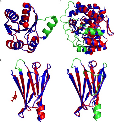
Figure 4.

Biological examples of order‐disorder transitions between protein conformations. The region suffering order‐disorder transition is coloured in red. (a) Protein DivK conformations present an order‐disorder transition between apo (PDB code: 1M5T_A ‐ color blue) and holo (PDB code: 1MB0_A ‐ color cyan) forms. These structures present a C‐apha RMSD of 0.345 A. (b) Enzyme dihydropteroate synthase (DHPS) shows an order‐desorden transition in the conformers PDB code: 3TYZ_B (color blue) and 3TZN (color cyan). These structures present a C‐alpha RMSD of 0.548 A. (c) Conformers of human Transthyretin (TTR), (c1) Apo form (PDB code: 1TTA_B ‐ color blue) and holo form (PDB code: 3D2T_B ‐ color cyan) in complex with diflunisal. (c2) Apo conformation of TTR (PDB code: 1TTA_B ‐ color blue) and apo form (PDB code: 3CBR_B ‐ color cyan) at pH 3.5.
