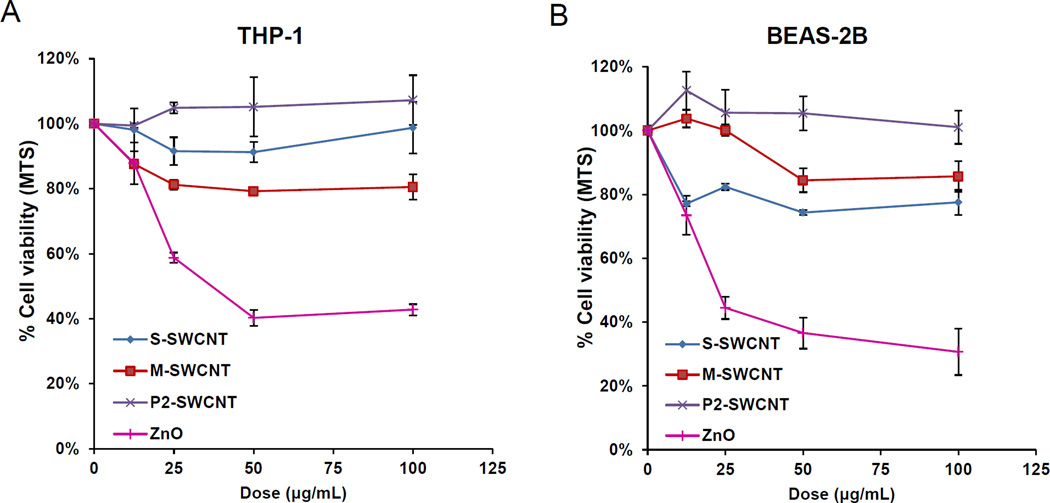
Figure 2. Cytotoxicity in THP-1 and BEAS-2B cells exposed to electronic-sorted SWNCTs.
Assessment of cytotoxicity of SWNCTs in (A) THP-1 and (B) BEAS-2B cells. Both cell types were grown in 96-well plates, followed by exposure to 12.5, 25, 50 and 100 µg/mL of each of the SWNCTs suspensions for 24 h. The media were subsequently washed with PBS and replaced with 120 µL aliquots of the MTS working solution. After incubation for 1 hour, the plates were centrifuged to collect the supernatants, and their absorbance read at 490 nm in a microplate reader (SpectroMax M5e, Molecular Devices, Sunnyvale, CA). All the MTS values were normalized according to the nontreated control, which was regarded as representing 100% cell viability. *p< 0.05 compared to control.

