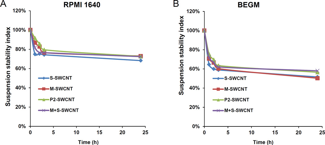
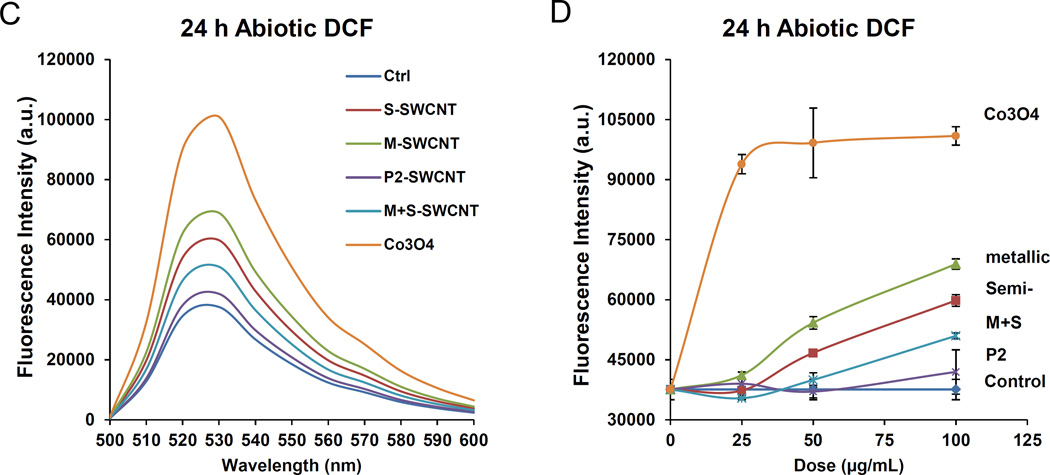
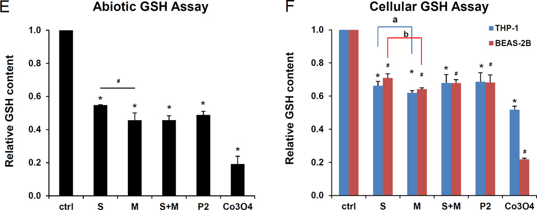
Figure 4. SWCNT suspension stability indices in cell culture media as well as assessment of the material pro-oxidant potential.
(A) The SWCNTs were added to RPMI 1640 at 50 µg/mL. The suspension stability index was determined by comparing the initial absorbance (t = 0) to the absorbance at 1, 2, 3, or 24 h. The absorbance measurements were carried out at λ = 550 nm in a SpectroMax M5e (Molecular Devices Corp., Sunnyvale, CA). (B) Similar assessment in BEGM. (C) DCF fluorescence spectroscopy: 29 µmol/L DCF was added to tube suspensions at 12.5–100 µg/mL for 24h, and emission spectra collected at 500−600 nm, with excitation at 490 nm. Both S- and M-SWCNTs showed significant enhancement of DCF fluorescence compared to the non-treated control. ROS production by Co3O4 NPs at 100 µg/mL was used as positive control. (D) Comparative expression of the fluorescence intensity at 528 nm demonstrated a proportionally bigger dose-dependent increase in fluorescence activity with metallic compared to semiconducting SWCNTs. (E) Abiotic GSH depletion: 62.5 µmol/L GSH was incubated with 100 µg/mL SWCNTs for 24 h, and the GSH level was determined by the luminescence-based GSH-Glo kit. * p < 0.05, compared to control; # p < 0.05 compared between S- and M-SWCNTs. (F) Cellular GSH depletion in THP-1 and BEAS-2B cells was determined by the same assay. Both cell types were exposed to SWCNTs at 100 µg/mL for 24 h. GSH abundance was calculated as the fractional luminescence intensity of treated vs. non-treated cells (in which the abundance was regarded as 1.0). * # p < 0.05, compared to control (* is for THP-1 and # for BEAS-2B cells); a and b p < 0.05 for comparing S- with M-SWCNTs (a is for THP-1 and b for BEAS-2B cells).


