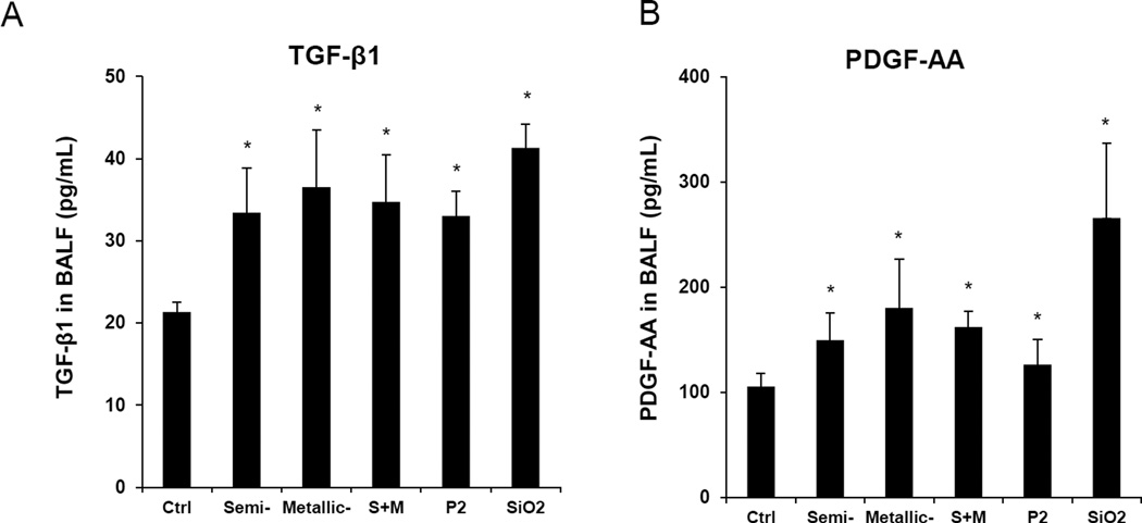
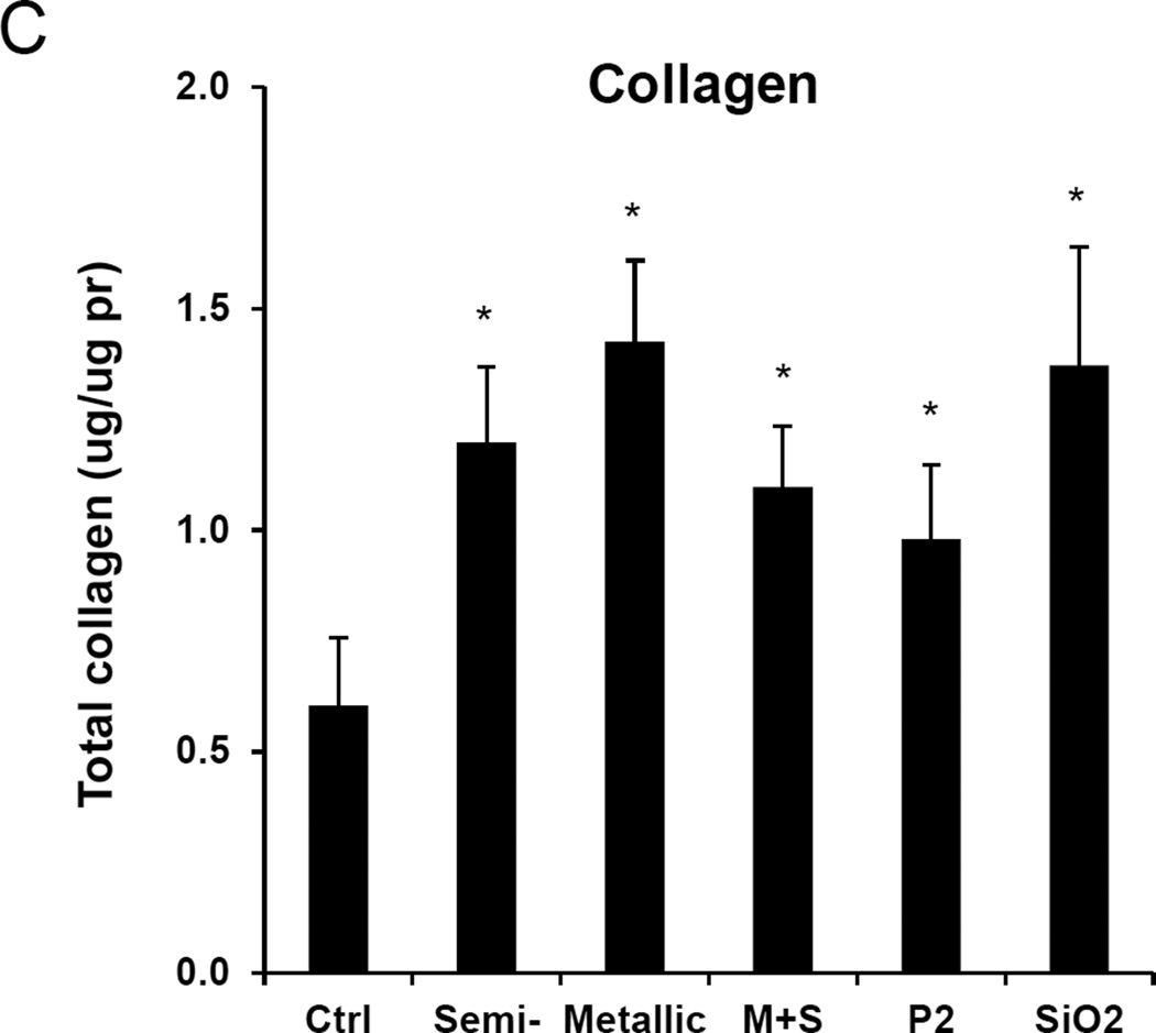
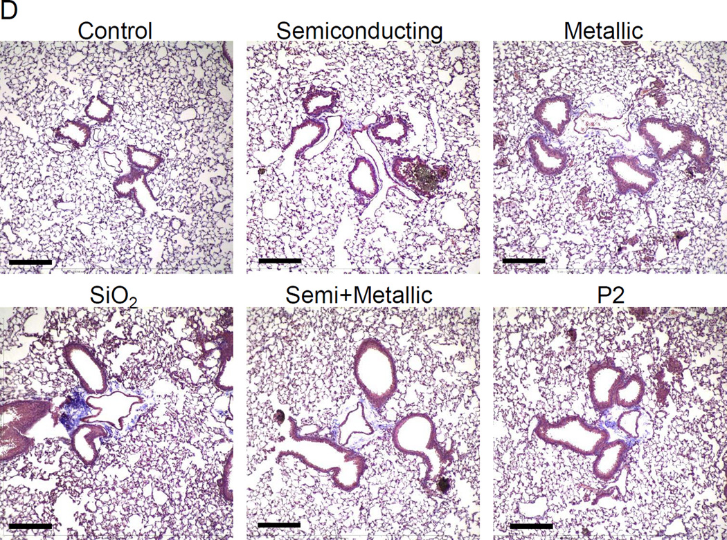
Figure 5. Assessment of the pro-fibrogenic effects of the electronic sorted SWNCTs in mice.
Anesthetized C57BL/6 mice were exposed to SWNCTs, delivered by one-time oropharyngeal aspiration of a 2.0 mg/kg bolus dose. Animals were euthanized after 21 d, and BALF was collected to determine (A) TGF-β1 and (B) PDGF-AA levels. (C) Assessment of total collagen content by a Sircol kit (Biocolor Ltd., Carrickfergus, UK). *p < 0.05 compared to control. (D) Visualization of collagen deposition in the lung, using Masson’s trichrome staining. Collagen deposition is shown as blue staining under 100 × magnification. Animals exposed to Quartz (QTZ) served as positive control.


